Send your good work in the knowledge base is simple. Use the form below
Students, graduate students, young scientists who use the knowledge base in their studies and work will be very grateful to you.
Introduction
1.Structure and functions of cell components
2.Mitotic cycle. Factors influencing mitotic activity
3. Criticism of ideas about fatality hereditary diseases
Literature
Introduction
The cage was discovered in the second half of the 17th century. The study of cells developed especially strongly in the second half of the 19th century in connection with the creation cell theory. Cellular level research has become the leading principle of the most important biological disciplines. A new section has taken shape in biology - cytology. The object of study of cytology is the cells of multicellular organisms, as well as organisms whose body is represented by a single cell. Cytology studies the structure, chemical composition, ways of their reproduction, and adaptive properties. This work will examine the structure and function of cell components.
Mitosis is an indirect cell division, as a result of which the original cell gives rise to two new ones that have exactly the same set of genes. The mitotic cycle is a set of processes as a result of which two new ones are formed from one cell; it covers the period of mitosis and part of the interphase. The goal of our work on this issue will also be to analyze the factors influencing mitotic activity.
Heredity is a fundamental property of all living things, which lies in the fact that all living organisms are capable of storing information about their structure and transmitting this information to other generations. Humanity has come a long way and not an easy path in order to correctly understand the causes and laws of heredity. The purpose of our work in the aspect of heredity will be to consider criticism of ideas about the fatality of hereditary diseases, which is especially relevant for today's medical research.
1.Structure and functions of cell components
The theoretical basis of cytology is cell theory. The cell theory was formulated in 1838 by T. Schwann, although the first two provisions of the cell theory belong to M. Schleiden, who studied plant cells. T. Schwann, a well-known specialist in the structure of animal cells, in 1838, based on data from the works of M. Schleiden and the results of his own research, made the following conclusions:
The cell is the smallest structural unit living organisms.
Cells are formed as a result of the activity of living organisms.
Animal and plant cells have more similarities than differences.
The cells of multicellular organisms are interconnected structurally and functionally.
Further study of its structure and life activity made it possible to learn a lot of new things about it. This was facilitated by the improvement of microscopic technology, research methods and the entry into cytology of many talented researchers. The structure of the nucleus was studied in detail, and a cytological analysis of such important biological processes as mitosis, meiosis, and fertilization was carried out. The microstructure of the cell itself became known. Cell organelles were discovered and described. Program cytological studies The 20th century set the task of clarifying and more accurately distinguishing the properties of the cell. From here Special attention began to be devoted to the study of the chemical composition of the cell and the mechanism by which the cell absorbs substances from the environment.
All these studies made it possible to multiply and expand the provisions of the cellular theory, the main postulates of which currently look like this:
The cell is the basic and structural unit of all living organisms
Cells are formed only from cells through division.
The cells of all organisms are similar in structure, chemical composition, basic physiological functions.
The cells of multicellular organisms form a single functional complex.
The cells of all living beings on earth can be divided into two fundamentally different types: nuclear (eukaryotic) and non-nuclear (prokaryotic). Prokaryotic cells are the oldest on our planet, these are the cells of bacteria and blue-green algae. They are characterized by the following features:
Lack of core.
Presence of circular DNA.
Multiple repetitions of identical genes in DNA.
Absence of self-dividing cell organelles: centrioles, mitochondria, plastids.
Cell division by amitosis (direct division).
Organisms of plants, fungi and animals are formed from eukaryotic cells. They appeared later than prokaryotes. They are characterized by such signs as:
The presence of a nucleus, where DNA molecules are always located. Some cells lose their nucleus for the second time (mammalian red blood cells and platelets).
DNA is always in the form of one or more strands, open at the ends.
Genes in each DNA molecule are usually not repeated.
Cells always contain self-dividing organelles that have their own DNA molecules: centrioles, mitochondria, plastids. The latter are found only in plant cells.
Cell division by mitosis (indirect division), as a result of which all genes are evenly distributed among new cells.
Eukaryotic cells are tens and hundreds of times larger than prokaryotic cells.
Let us consider in more detail the structure of a eukaryotic cell.
A cell has a membrane, cytoplasm and nucleus.
The membrane is a cell organelle with a four-layer structure. The outer and inner layers are protein. Between them lie two layers of fat-like substances - lipoids. One of the ends of the lipid molecule has pronounced hydrophobic properties. In the membrane, all lipoids are located so that with their hydrophobic ends each layer is oriented in the opposite direction from the other. IN different places The cell membrane contains special large protein molecules that occupy its entire thickness. The membranes of many cells are covered on the outside with additional protective membranes, consisting of either carbohydrates (for example, cellulose in plant cells) or complex substances - glucoproteins (pellicles of ciliates and flagellates). The health of the cell and its lifespan largely depend on the condition of the membrane.
Completely permeable to water. The membrane always allows water to pass into or out of the cell, depending on where the water concentration is greater. This movement of a substance from an area of high concentration to an area of lower concentration is called diffusion. Diffusion of matter does not require energy.
Selective conductivity of solutes:
Negatively charged particles penetrate the membrane faster and more easily.
Fat-soluble substances penetrate the membrane more easily than water-soluble substances.
Small molecules penetrate the membrane more easily than large ones.
Active transport of substances. Some substances are able to penetrate the membrane in the direction opposite to their diffusion, that is, from a place of low to a place of higher high concentration. By active transport, excess sodium, hydrogen and chlorine ions are constantly removed from the cell. On the contrary, phosphates, glucose, and amino acids actively penetrate into the cytoplasm. Active transport always involves energy consumption.
The membrane is regularly restored as a result of the work of special organelles that synthesize membrane vacuoles. Many membranes not covered dense shells, are capable of forming temporary outgrowths called pseudopods (pseudopodia).
Membrane functions:
Phagocytosis is the capture of solid food particles by pseudopods. As a result, a digestive vacuole is formed, floating in the cytoplasm.
Pinocytosis is the absorption of solutes.
Protective. The membrane protects the cell from the penetration of foreign, dangerous substances.
Respiratory. Oxygen enters the cell through the membrane and carbon dioxide is released.
Homeostatic. Homeostasis is the ability to maintain a relatively constant composition. Due to its properties (selective absorption of substances and active transport), the membrane provides the cell with a constant composition.
Integrative. Cells communicate with each other using membranes. Through the membrane, one cell can transmit various information to another cell. This information can be transmitted both using electrical impulses and using chemical substances(hormones, mediators).
Cytoplasm is cell sap, cell fluid. Contains water, inorganic and organic substances dissolved in it, as well as various separate structures called organelles:
Ribosomes are cell organelles consisting of two particles, large and small. Each particle is formed by proteins and ribosomal RNA. Ribosomes carry out protein synthesis. Synthesized in the nucleus.
The endoplasmic reticulum (ER) is a membrane organelle of a cell, representing numerous channels and cavities of membranes, similar in structure to the cell membrane. According to structure and function, it is divided into two types: rough ER - contains ribosomes on the surface and is the site of protein synthesis; smooth ER - does not contain ribosomes, is the site of synthesis of carbohydrates, lipoids and fats. Outside, the EPS is in contact with the cell membrane, inside - with the nuclear membrane.
Golgi apparatus - by location it is a part of the endoplasmic reticulum. Has a membrane structure. It looks like a cluster of numerous sacs, cavities, and vacuoles. Performs many functions:
Brings proteins to their final working form, some proteins into large protein complexes, attaches essential metal ions to some proteins.
Forms membrane vesicles, which, leaving the Golgi complex, either restore the cell membrane or turn into lysosomes.
Lysosomes are membrane organelles of the cell, representing microscopic vesicles filled with digestive enzymes. Perform digestive and protective functions. They can adhere to the digestive vacuole, pouring into it digestive enzymes. When a cell comes into contact with a foreign substance or with a foreign cell, lysosomes adhere to the cell membrane, releasing their enzymes outside the cell. Lysosome enzymes can also take part in the programmed death of one's own cell.
Mitochondria are membrane-bound, self-dividing organelles. They are formed by two layers of membranes: an outer smooth one and an inner one, which has numerous projections into the mitochondria. Such outgrowths of the inner membrane are called cristae. The process of oxidation of lactic acid occurs in them, as a result of which energy is released, stored in the form of ATP (oxidative phosphorylation). Therefore, the most important function of mitochondria is energy. Mitochondria have their own DNA molecules, which are no different in structure from bacterial DNA. Mitochondria, like bacteria, reproduce by direct fission.
Plastids are membrane-bound, self-dividing cell organelles. Unlike all the organelles discussed above, plastids are found only in plant cells. Their structure resembles mitochondria: they are formed by two membranes, the outer smooth and the inner, which forms numerous flat outgrowths - thylakoids. All thylakoids are arranged in stacks like stacks of coins. Each stack is called a face. Between the grana there is an internal fluid of the plastid called stroma. It contains its own DNA, which is similar in structure to bacterial DNA. Plastids reproduce like bacteria by direct division.
Centrioles are self-dividing organelles of animal cells and some lower plants. Each centriole consists of a short hollow cylinder, the walls of which are formed by microtubules located along the axis of the cylinder. Centrioles contain proteins and a small amount of RNA. There are two pairs of centrioles in the cell.
2.Mitotic cycle. Factors influencing mitotic activity
The mitotic cycle is a set of processes as a result of which two new ones are formed from one cell. The mitotic cycle covers the period of mitosis and part of interphase. -- the period between divisions, when preparation for the next mitosis occurs. The mitotic cycle is part of the cell's life cycle; in rapidly dividing cell populations (for example, in blastomeres of a cleaving egg), the mitotic cycle almost coincides with life cycle cells.
Mitosis is an indirect cell division, as a result of which the original cell gives rise to two new ones that have exactly the same set of genes.
Mitosis lasts 1-2 hours and occurs in four phases, of which the first and last are the longest.
Phases of mitosis.
Prophase. Condensation of chromatin threads is observed, that is, their packaging. Thickened chromosomes, clearly visible under a light microscope (with special staining), are formed. The synthesis of RNA and proteins ends. The shell of the core is destroyed. A fission spindle is formed.
Metaphase. All chromosomes move to the center of the cell, located along its equator. Each chromosome consists of two clearly distinguishable daughter chromatids formed by daughter DNA resulting from reduplication of the mother. Any pair of daughter chromatids is connected to each other by a thin intercept called a centromere. This is a section of maternal DNA in which reduplication has not yet taken place. Each centromere has its own spindle filament attached.
Anaphase. Daughter chromatids are separated from each other as a result of centromere reduplication and quickly diverge to opposite poles of the cell. Now each pole has its own set of chromatids. Both of these sets contain the same genes, since all daughter chromatids formed during the reduplication of maternal DNA are copies of each other.
Telophase. At the cell poles, chromatids unwind into chromatin filaments. The synthesis of RNA and proteins resumes. Around each set of daughter chromatids, their own nuclear membranes are formed. The cage is laced along the equator. Two new cells are formed.
As a result of mitotic division, two cells appear genetically absolutely identical. This is only possible through two processes:
DNA reduplication, which is based on the principle of complementarity.
the divergence of each pair of daughter chromatids into new cells.
Mitotic cell division occurs:
at asexual reproduction plants, mushrooms and animals,
during the embryonic and postembryonic development of all multicellular organisms from a fertilized egg,
during wound healing, formation of blood cells, growth of skin and intestinal epithelial cells, and other processes.
As a result of irradiation of a very large number of cells of the same type, it was established that when exposed to different types radiation, the duration of reversible inhibition of cell division and the percentage of cells in which division has completely stopped increase as the radiation dose increases. As the radiation dose increases, an increasing number of cells lose their ability to reproduce, or at least their division process temporarily stops. One of the indicators of a violation of this ability of cells to reproduce in both single-celled and tissue cells higher organisms is the emergence of giant cell forms.
Some radiation-biochemical changes appear after exposure to relatively small doses, while other changes occur only as a result of exposure to medium or high doses radiation. Among the metabolic disorders that occur when exposed to ionizing radiation, the first place should be put on the violation of the most radiosensitive substrate—nucleic acids. Radiation injuries in the form of inhibition of nucleic acid synthesis cannot be considered as a direct cause of inhibition of cell division or chromosome breakage, which can lead to their gross morphological disorders, determined during mitosis after irradiation. Violations of other types of metabolism, for example carbohydrate metabolism, give the right to talk about its very low radio sensitivity. Changes carbohydrate metabolism after irradiation, in particular, the inhibition of anaerobic glycolysis becomes noticeable, as a rule, only after exposure to doses of about 5,000-20,000 rubles; disruption of cellular respiration is usually observed as a result of exposure to even higher doses - from 20,000 to 100,000 r.
When exposed to low doses of radiation, inhibition of cell division is observed. At large doses cells finally lose their ability to reproduce. Temporary inhibition of mitosis and complete sterility cannot be caused by a single mechanism, despite the fact that both of these phenomena at first glance may seem quite related.
From the quality of radiation, except functional changes, also depend certain types radiation chromosomal aberrations. In cell populations with mitotic cell division after irradiation, there is first a short-term increase in the frequency of mitoses, and then a drop to a certain minimum value.
The primary and secondary effects of radiation are characterized by certain types of chromosomal changes.
The mechanism of chromosomal changes during the primary and secondary effect is different. Chromosomal changes typical of the primary effect occur mainly in those cells that had mitotic activity and were in the metaphase stage during irradiation. A certain number of these cells exhibit mitoses, the frequency of which decreases as a result of irradiation. In other mitotically dividing cells that have reached or passed the metaphase stage, mitoses continue, but at a slower pace.
A certain radiation is associated with cell division (mitosis), which was discovered and measured by A.G. Gurvich. He called it “mitogenetic.” It was found that if other cells are exposed to this radiation, then their mitosis increases, that is, their growth is stimulated.
3. Criticism of ideas about the fatality of hereditary diseases
Until recently, even among doctors, the prevailing opinion was that hereditary diseases are fatal and that their prevention and treatment are impossible. Today, treatment methods have already been found for some diseases. Phenylketonuria is observed on average in every 10 thousand newborns. As a result of the absence in the body of the enzyme that controls the conversion of the amino acid phenylalanine into tyrosine, the concentration of phenylalanine increases tens of times. Part of it is excreted in the urine, and the rest is converted into phenylpyruvic, phenylacetic, phenyllactic acid and other substances. This causes a number of secondary biochemical changes, as a result of which brain maturation is disrupted. Deviations in mental development the child becomes noticeable only after 6 months of age. Most children suffering from phenylketonuria grew up mentally retarded. Now this metabolic disorder is eliminated by a protein-free diet, which the child is on until 6-8 years of age. Protein substances are given only in the form of special preparations from which phenylalanine has been removed. Diagnosis of the disease is quite simple: it is based on a positive qualitative reaction of urine with phenylpyruvic acid.
Another hereditary disease - galactosemia - often manifests itself in the first days of a child’s life with vomiting, severe lethargy, hypotension, jaundice, and convulsions. If the disease develops gradually, the main symptoms appear somewhat later. These include cataracts, mental retardation and chronic liver damage - hepatitis. Patients have increased galactose content ( milk sugar), and glucose levels are reduced. If the diagnosis is made in a timely manner and dairy products are excluded from the child’s diet, the child’s development is normal.
We have given only two examples. There are much more hereditary diseases. According to the World Health Organization, about 4% of newborns suffer from some kind of genetic defect. But to them we must add those children in whom the disease does not appear immediately after birth, but at a later age. Therefore, the further development of medical genetics and the dissemination of genetic knowledge not only among doctors, but also among the population is an important task. Not divine predestination, but real reasons form the basis of each type of hereditary pathology. The fight against these diseases is carried out in two ways. The first way is a targeted change in environmental conditions, making the development of the disease impossible. The second is prevention through medical and genetic counseling of the population.
Hereditary metabolic disorders are corrected special diet: elimination from food of substances indigestible by the body or, on the contrary, introduction of missing ones. Many defects of the organs of speech, hearing, and vision are corrected surgically. Not knowing true reasons the birth of children with congenital deformities, people often regarded this as “God’s punishment for sins” or a harbinger of serious misfortunes. J. W. Ballantyne in his book "Teratological Records of the Chaldeans" (1894) gives examples different interpretations and predictions related to the birth of freaks: "If any woman gives birth to a child who has no nostrils, the country will be threatened with misfortune, and her husband's house will be destroyed. If any woman gives birth to a child who has no nose, disaster will befall the country , and the owner of the house will die. If any woman gives birth to a child who does not have a penis, the owner of the house will reap a rich harvest from the fields. If a woman gives birth to a child whose sex is not clearly indicated, disasters and misfortunes will befall the country, and her husband will accompany misfortune."
In the origin of neuroses and reactive psychoses play a major role mental trauma, which sometimes only provoke hereditary predisposition to illness. In origin mental illness combination plays a role causal factors With individual characteristics person. For example, not all people suffering from syphilis develop syphilitic psychosis, and only a small number of patients with cerebral atherosclerosis develop dementia or hallucinatory-delusional psychosis.
Today, many hereditary diseases of the unborn child are diagnosed at the beginning of pregnancy by examining amniotic amniotic fluid. This lets you start timely treatment in cases where it is possible and appropriate, or to terminate the pregnancy in order to prevent the birth of a defective child. Parents must be informed about the degree of misfortune that threatens them in order to make the right decision.
conclusions
Thus, all organisms consist of the same parts - cells; they are formed and grow according to the same laws. The general principle of development for the elementary parts of the body is the formation of cells. Each cell within certain boundaries is an individual, a kind of independent whole. But these individuals act together, so that a harmonious whole fabric emerges. All tissues are made up of cells. The processes occurring in plant cells boil down to the following: the emergence of new cells, an increase in cell size, changes in cellular contents and thickening of the cell wall.
Thanks to the creation of the cell theory, it became clear that the cell is the most important component of all living organisms. Tissues and organs are made up of cells. Development always begins with a single cell, and therefore we can say that it represents the precursor of a multicellular organism. Structure and functions cellular components were considered in this work.
As we can see, the ability to divide - most important property cells. Without division, it is impossible to imagine an increase in the number of single-celled creatures, the development of a complex multicellular organism from one fertilized egg, the renewal of cells, tissues and even organs lost during the life of the organism.
Cell division occurs in stages. At each stage of division certain processes occur. They lead to the doubling of genetic material (DNA synthesis) and its distribution between daughter cells. The period of cell life from one division to the next is called the cell cycle. During mitosis, the cell goes through a series of successive phases, as a result of which each daughter cell receives the same set of chromosomes as was in the mother cell.
There are 4 phases of mitosis: prophase, metaphase, anaphase and telophase. Among the factors influencing mitotic activity, we paid special attention to radiation-biochemical influences.
The opinion about the fatality of hereditary diseases, the impossibility of their prevention and treatment, which prevailed even among doctors until recently, is increasingly being criticized. And today, treatment methods have already been found for some diseases.
In addition, at present, many hereditary diseases of the unborn child can be diagnosed at the beginning of pregnancy by examining amniotic amniotic fluid, which makes it possible to begin timely treatment, if appropriate, or to terminate the pregnancy in order to avoid the birth of a defective child.
Literature
1. Adamchik M.V. - World Encyclopedia. Biology / Ed. Anikeev V.I. - M.: Modern writer, 2006.
2. Kalyuzhny V.G. Handbook of biology. (Series "Textbooks and teaching aids") - Rostov-on-Don, Phoenix, 2004.
3. Gilyarov M.S. Biology. Large encyclopedic dictionary. - 3rd ed. - (Gold Fund). - M.: Great Russian Encyclopedia, 2005.
4.B. Smelova. Cell structure. Newspaper "Biology". - No. 36/2001.
5.P. Wallet. The doctrine of the plant cell. - http://bio.1september.ru/article.php?ID=200204304
6.I. Bologova. A cell is a structural and functional unit of living things. - http://bio.1september.ru/article.php?ID=200200603
7. Grekova T.I. Atheism and medicine. - http://lib.metromir.ru/author5836
8. N. Green, W. Stout, D. Taylor. Biology. M.: Mir, 1996.
Similar documents
History and main stages of research into the cell, its structure and components. The content and significance of cell theory, outstanding scientists who contributed to its development. Symbiotic theory (chloroplasts and mitochondria). The origins of a eukaryotic cell.
presentation, added 04/20/2016
Methods for studying cells, their dependence on the type of microscope lens. Provisions of the cell theory. Animal cells and plant origin. Phagocytosis - absorption by a cell from environment dense particles. Approaches to the treatment of hereditary diseases.
presentation, added 09/12/2014
The nucleus of a eukaryotic cell. Cells that have more than two sets of chromosomes. The process of division in eukaryotes. United pairs of homologous chromosomes. Ontogenesis of a plant cell. The process of cell separation as a result of destruction of the median lamina.
abstract, added 01/28/2011
Periods and phases of the cell cycle. The sequential passage of a cell through cycle periods without skipping or returning to previous stages. Division of the original cell into two daughter cells. Cyclins and cyclin-dependent kinases; eukaryotic cell division; mitosis.
test, added 11/21/2009
Authors of the creation of cell theory. Features of archaea and cyanobacteria. Phylogeny of living organisms. The structure of a eukaryotic cell. Membrane mobility and fluidity. Functions of the Golgi apparatus. Symbiotic theory of the origin of semi-autonomous organelles.
presentation, added 04/14/2014
Elements of cell structure and their characteristics. Functions of the membrane, nucleus, cytoplasm, cell center, ribosome, endoplasmic reticulum, Golgi complex, lysosomes, mitochondria and plastids. Differences in the cell structure of representatives of different kingdoms of organisms.
presentation, added 11/26/2013
Types of cell damage. Stages of chronic cell damage. Types of cell death. Necrosis and apoptosis. Pathogenesis of damage cell membranes. Highly specialized cells with high level intracellular regeneration. Conditions of connective tissue.
presentation, added 11/03/2013
Elementary genetic and structural-functional biological system. Cell theory. Types cellular organization. Features of the structure of a prokaryotic cell. Principles of eukaryotic cell organization. Hereditary apparatus of cells.
test, added 12/22/2014
Structure animal cell. Basic provisions of cell theory, the concept of prokaryotes and eukaryotes. The structure of the cytoplasm and the endoplasmic reticulum. Human chromosome set. Methods of cell division (amitosis, mitosis and meiosis) and its chemical composition.
presentation, added 10/09/2013
Compartmentation in the organization of the eukaryotic cell. Linear dimensions of a eukaryotic cell. Nuclear-cytoplasmic ratio. Various shapes chondrioma. Mitochondrial system of cardiomyocytes. Signs of mitochondrial diseases in humans.
Cell. Structure and interaction of cellular elements. Elements of cellular pathology.
(Prof. Semenov V.V., associate Koshpaeva E.S., associate Kolochkova E.V. took part in the preparation of the lecture)
ISSUES CONSIDERED.
1. INTRODUCTION
2. Structural Components cells.
2.1. The plasma membrane and its role in the life of the cell.
2.1.1. The structure of the plasma membrane.
2.1.2. Functions of the plasma membrane.
A. Transport function.
b. Transferring information via plasma membrane.
2.1.3. Participation of membrane components (lipids, proteins and glycoproteins) in pathological processes.
A. Lipid peroxidation.
b. Phospholipase activity.
V. Glycoproteins and the tumor process
2.2. Endoplasmic reticulum.
2.3. Golgi complex.
2.4. Lysosomes.
2.5. Ribosomes.
2.6. Mitochondria.
2.7. Cell center.
2.8. The nucleus is the cell control system.
2.8.1. Nuclear envelope.
2.8.2. Nucleoplasm (karyolymph, nuclear juice).
2.8.3. Morpho-functional characteristics and classification of chromosomes.
A. Chromatin – euchromatin and heterochromatin.
b. Nucleosome.
2.8.4. Nucleolus.
2.8.5. Nuclear matrix.
2.9. Cytoplasmic inclusions.
A. Trophic inclusions.
b. Secretory.
V. Pigmented.
g. Excretory.
In this lecture we will move away from the traditional presentation of the morpho-functional (structure and function) features of the cell, which were characteristic of school teaching methods. We will focus on the functional side of the behavior of the cell and its elements. In doing so, we proceed from three important in medical biology principles. Firstly, the cell, as an integral system, is not located in the external natural environment, but inside the entire organism , in a peculiar liquid intercellular environment. And this is no coincidence. The evolution of living things began in the aquatic environment. Almost all the main morphological formations of the cell have evolved in an aquatic environment. They are adapted to it. After the transition to land, into a gaseous environment, it was necessary to significantly change the entire organization of living things adapted to the aquatic environment. Now it is difficult to guess for what reasons the evolution in a gas environment in organisms that reached land could not completely eliminate all the adaptations that organisms received while in a liquid environment. Perhaps evolution in a gaseous environment required such radical changes in living systems adapted to an aquatic environment that they were simply incompatible with life. According to another option, the evolution of organisms adapted to an aquatic environment was impossible in a gas environment. Perhaps there were some other reasons, but in any case, Nature made a compromise - the organisms that came to land contained within themselves the environment of the primary ocean to which they were adapted. In fact, blood, lymph, and intercellular fluid in their composition resemble the cradle of our development, the primary ocean in which the first stage of our evolution took place. The cell, being inside the body, has virtually no contact with the external environment; all its activities take place in intercellular fluid, which ensures not only the existence of the cell, but is also the initiator of the restructuring of its metabolism. This restructuring transfers the cell to a new mode of life activity, to a different functional state. However, such a transition is possible as a result of the second important principle - the structure and functions of the subcellular organelles of the cell are not strictly determined, they are plastic, capable of changing within certain limits. And since intracellular elements are involved in various biochemical processes, any changes in the structure of the cell will certainly lead to a change in the functions performed by this structure. What is characteristic of living things is that the same cellular element can perform several functions. For example, this is well illustrated below when listing the functions of, for example, organelles. And finally, the third principle must be remembered:- all intracellular elements and processes represent a single interconnected system, this set of elements and processes can be represented as a kind of network in which a change in one cell or node leads to a change in the entire intracellular organization and its function. This principle has great importance in medicine, because sometimes the changes that occur are so strong that the coherent internal organization of the cell is disrupted. In this case, the processes responsible for self-regulation, adaptation and others cellular functions can become an arena for the development of pathology, first at the level of intracellular elementary structures and processes, then at the level of pathology of the entire cell, as an elementary self-regulating living system, and then at the level of cellular formations united by a final function.
2. Structural components of a eukaryotic cell
On outside cells (Fig. 1) there is an outer plasma membrane that separates the cell from external environment. Below it are the cytoplasm and the nucleus. The nucleus is not always located in the center of the cell. In cases where intensive work is carried out in one part of the cell, for example. there is an active absorption process nutrients with the expenditure of energy, the nucleus is shifted to the opposite “non-working” part of the cell, and mitochondria are concentrated in the “working” part. The cytoplasm and nucleus, in turn, consist of several components, which are presented in Figure 1.
The structural organization of the cell is based on membrane principle. This means that membranes constitute an essential part of the structure of the cell. They separate the cell from the surrounding intercellular environment and divide the cell into separate compartments - compartments. These isolated areas contain specific metabolic processes. This differentiation in evolution increased the organization of the cell, but it did not end there. Over time some biochemical processes in compartments have become so specific that there is a need to create more isolated, almost autonomous structures - organelles. Membranous organelles include mitochondria, plastids, lysosomes, endoplasmic reticulum and Golgi complex. These bridgeheads house vital processes for the body. At the same time, structures were created that do not have a membrane membrane, but also perform certain cellular functions - ribosomes, cell center, etc. At the same time, the cytoplasm of the cell remains isolated by the membrane of the endoplasmic reticulum, although not completely, into separate compartments in which they are also localized certain parts of cellular metabolism, for example, glycolysis. All biomembranes are built approximately the same, so we will consider the structure of only the plasma membrane.

http://xn--d1aacnkch5m.xn--p1ai/14-bez-rubriki/images/image004.gif" border="0" width="104" height="55 src=">
http://xn--d1aacnkch5m.xn--p1ai/14-bez-rubriki/images/image006.gif" border="0" alt=" Signature:" width="126" height="64 src=">http://xn--d1aacnkch5m.xn--p1ai/14-bez-rubriki/images/image008.gif" border="0" width="204" height="48 src=">!}
http://xn--d1aacnkch5m.xn--p1ai/14-bez-rubriki/images/image010.gif" border="0" width="12" height="87 src=">http://xn- -d1aacnkch5m.xn--p1ai/14-bez-rubriki/images/image012.gif" border="0" width="12" height="99 src=">http://xn--d1aacnkch5m.xn-- p1ai/14-bez-rubriki/images/image014.gif" border="0" width="12" height="13 src=">http://xn--d1aacnkch5m.xn--p1ai/14-bez- rubriki/images/image016.gif" border="0" width="12" height="81 src=">http://xn--d1aacnkch5m.xn--p1ai/14-bez-rubriki/images/image011. gif" border="0" width="12" height="27 src=">
Lipids Nuclear juice
Proteins Hyaloplasm
Organelles Chromosomes
Polysaccharides Inclusions
Nuclear membrane
Rice. 1. Overall plan structure of a eukaryotic cell.
2.1. Plasmatic membrane, its role in cell life
2.1.1. The structure of the plasma membrane.
The membrane consists of two layers of lipid molecules (bilayer), into which proteins are embedded. Some lipid and protein molecules have carbohydrates attached to them. There are not many of them. The thickness of the membrane is about 10 nm (0.00001 mm). The main part of the membrane is a continuous layer of phospholipid molecules. In that liquid layer immersed in molecules of protein molecules of different structure and functions. Proteins do not completely cover the lipid bilayer, but are located in it individually or in groups. Overall this reminds me mosaic(Fig. 2. B. C). In this regard, the currently accepted membrane model is called liquid mosaic. Proteins are capable move along the lipid layer. The molecules of the lipid layer also move. It is clear that the movement of membrane molecules changes the physicochemical characteristics of the latter, and this, in turn, changes the functional characteristics of the membrane. It should be noted that the plasma membrane of most cells does not have the shape of a perfect sphere. On the contrary, it has many protrusions and depressions that constantly change their shape and size. Received in Lately the results made some adjustments to the theory of membrane structure. It has been shown that not all membrane proteins are capable of movement, and some membrane regions differ in structure from the classical lipid bilayer.
The phospholipid molecule looks like a head with two tails (Fig. 2 A). The head (glycerol) is soluble in water, hydrophilic, the tails (fatty acids) are insoluble in water, hydrophobic. Therefore, being in water, molecules spontaneously occupy a certain position in relation to the aqueous phase. Starting from water molecules, the tails are located deep in the lipid layer, and the water-soluble heads are facing the external and internal aqueous environment (Fig. 2. B). The lipid bilayer is called the matrix. Of particular note is the presence in cell membranes of lipids, the tail of which contains fatty acids that have double bonds in their structure located through the CH2 group (– CH = CH – CH = CH – CH –). Such fatty acids are called unsaturated. These acids are most susceptible to the effects of reactive oxygen species (ROS), which are constantly present in the body of all living things. Their number especially increases with various diseases. This may lead to adverse consequences, which we will talk about below.
Proteins embedded in the matrix (Fig. 2 B) are located differently. Some on the inner and outer surface of the lipid layer are near-membrane or surface proteins, others are semi-immersed in the membrane - semi-integral proteins, and others permeate the entire membrane - integral proteins. Semi-integral and integral proteins are usually combined under the same term, intrinsic proteins, because they are difficult to distinguish from each other. Integral proteins are most often found in membranes. They can be represented either by one molecule and perform any one function, or by a group (ensemble) of proteins. Each member of the ensemble performs a strictly defined role. These complexes also perform one or more terminal functions.
A B BMsoNormalTable" style="border-collapse: collapse; border: none; margin-left: 6.75pt; margin-right: 6.75pt;" align="left">
Phospholipid
1-head,
2-tail
Lipid bilayer
(matrix)
The location of protein molecules relative to the lipid layer: 1 - near-membrane proteins, 2 - semi-submerged protein, 3 - integral protein
Rice. 2. Schematic structure of the plasma membrane.
It should be noted that integral and surface enzyme proteins functioning in the membrane often change their position. In some cases, it is difficult to determine which type (surface or integral) a particular membrane protein belongs to. For example, the enzyme phospholipase A, deposited on the membrane, is a surface protein, but then it is activated, becomes an integral protein and, interacting with bilayer lipids, forms from them arachidonic acid(Fig. 3). The latter leaves the membrane and is converted into other active compounds that participate in the development of various pathological processes.
Na+, K+- ATPase Adrenoreceptor Adenylate cyclase
http://xn--d1aacnkch5m.xn--p1ai/14-bez-rubriki/images/image020.gif" border="0" width="12" height="79 src=">http://xn- -d1aacnkch5m.xn--p1ai/14-bez-rubriki/images/image022.jpg" border="0" width="576" height="193 src="> Ca2+-ATPase
http://xn--d1aacnkch5m.xn--p1ai/14-bez-rubriki/images/image030.gif" border="0" width="12" height="39 src="> cAMP
Na+, K+ Ca2+ Arachidonic
acid G-protein Glycogen
Rice. 3. Hypothetical scheme of localization of some membrane proteins
On the contrary, proteins involved in the movement of substances across the membrane, for example, proteins involved in the facilitated diffusion of Na+, K+-ATPase or Ca2+-ATPase, as a rule, do not change their position, functioning as integral proteins (Fig. 3 ). And finally, as we have already said, the membrane can contain complex complexes of several proteins connected into a single ensemble with one task. Such complexes include proteins that take part in conducting an information signal through the membrane (Fig. 3). The latter includes a complex containing three proteins - adrenergic receptor, G-protein and adenylate cyclase. All these proteins have essential in normal cell activity and in pathology. We will talk about this below.
In addition to lipids and proteins, the membrane contains carbohydrates, but they are not located as independent components, but as components of lipids (glycolipids) or proteins (glycoproteins). Carbohydrates are located on the outer surface of the plasmalemma.
2.1.2. Functions of the plasma membrane.
The plasma membrane has many functions. Let's list the most important ones.
· Transfer of substances across the membrane. Through the membrane, substances are transported to both sides of the membrane.
· Transfer of information through the membrane. On the membrane, information from outside is perceived, converted and transmitted into or out of the cell. Membrane receptors play a significant role in this.
· Protective role. a) protects the contents of the cell from mechanical damage, chemical reagents and biological aggression, for example from the penetration of viruses, etc.;
b) in multicellular organism plasma membrane receptors form the immune status of the body;
c) in a multicellular organism, the membrane ensures the phagocytosis reaction.
- Enzymatic - membranes contain various enzymes (for example, phospholipase A, etc.), which carry out whole line enzymatic reactions. Glycoproteins and glycolipids on the cytoplasmic membrane make contact with the membranes of other cells.
Let's look at some of the listed functions in more detail.
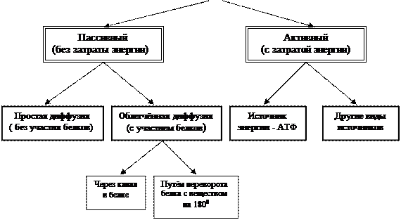
A. Transport function. Movement occurs through the membrane into and out of the cell. various substances, including medications. Depending on the size of the molecules transported through the membrane, two types of transport are distinguished: without violating the integrity of the membrane and with violating the integrity of the membrane. The first type of transport can be carried out in two ways - without energy consumption (passive transport) and with energy consumption (active transport) (see Fig. 4). Passive transfer occurs due to diffusion along an electrochemical gradient as a result of Brownian motion of atoms and molecules. This type of transport can be carried out directly through the lipid layer, without any participation of proteins and carbohydrates, or with the help of special proteins - translocases. The lipid layer mainly transports molecules of substances that are soluble in fats, and small uncharged or weakly charged molecules, such as water, oxygen, carbon dioxide, nitrogen, urea, fatty acids, as well as many organic compounds (for example, drugs) that are highly soluble in fats Translocases can transport a substance across membranes towards its lower concentration, without expending energy, using two different mechanisms - through a channel that runs inside the protein, or by connecting the part of the protein protruding from the membrane with the substance, rotating the complex by 1800 and detaching the substance from squirrel. The diffusion of substances through a membrane with the participation of proteins is important in that it occurs much faster simple diffusion through the lipid layer without the participation of proteins. Therefore, diffusion in which translocases take part is called facilitated diffusion. According to this principle, some ions (for example, chlorine ion) and polar molecules, as well as glucose, are transported into the cell.
Active transport of substances across a membrane is characterized by three properties:
1. Active transport occurs against a concentration gradient.
2. Carried out by a transporter protein.
3. Comes with energy consumption.
Energy during active transport of substances is necessary in order to transport a substance against its concentration gradient. Active transfer systems are often called membrane pumps. Energy in these systems can be obtained from various sources, most often such a source is ATP. The cleavage of phosphate bonds in ATP is carried out by the integral protein-enzyme ATPase. Therefore, this enzyme is found in the membrane of many cells in the form of an integral protein. The important thing is that this enzyme not only releases energy from ATP, but also moves the substance. Therefore, the active transport system most often consists of one protein - ATPase, which receives energy and moves the substance. In other words, the process of movement and energy supply in ATPase are coupled. Depending on what substances ATPase pumps, the pumps are called or Na+,K+- ATPase orCa2+-ATPase. The former regulate the content of sodium and potassium in the cell, the latter regulate calcium (this type of pump is most often located on the EPS channels). Let us immediately note the important medical workers fact: for the successful operation of the potassium-sodium pump, the cell spends about 30% basal metabolic energy. This is a very large volume. This energy is spent on maintaining certain concentrations of sodium and potassium in the cell and intercellular space; - the cell contains more potassium than the intercellular space, sodium, on the contrary, more in the intercellular space than in the cell. This distribution, far from osmotic equilibrium, provides the most optimal mode cell work.
 |
Rice. 4. Classification of types of transport of substances through the membrane.
By active transfer, inorganic ions, amino acids and sugars, and almost all medicinal substances with polar molecules move through the membrane - para-aminobenzoic acid, sulfonamides, iodine, cardiac glycosides, B vitamins, corticosteroid hormones, etc.
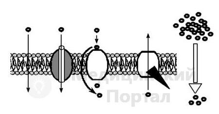
To clearly illustrate the process of transfer of substances through the membrane, we present (with minor changes) Figure 5 taken from the book “Molecular Biology of the Cell” (1983) by B. Alberts and other scientists considered leaders in the development of the theory
 http://xn--d1aacnkch5m.xn--p1ai/14-bez-rubriki/images/image036.gif" border="0" width="38" height="19 src=">http://xn- -d1aacnkch5m.xn--p1ai/14-bez-rubriki/images/image038.gif" border="0" width="33" height="16 src="> protein transporter
Passive transport Active transport
Figure 5. Many small, uncharged molecules pass freely through the lipid bilayer. Charged molecules, large uncharged molecules, and some small uncharged molecules pass through membranes through channels or pores or with the help of specific transporter proteins. Passive transport is always directed against the electrochemical gradient towards the establishment of equilibrium. Active transport occurs against an electrochemical gradient and requires energy expenditure.
transmembrane transport, reflects the main types of transfer of substances across the membrane. It should be noted that the proteins involved in transmembrane transport belong to integral proteins and are most often represented by one complex protein.
The transfer of high molecular weight protein molecules and other large molecules through the membrane into the cell is carried out by endocytosis (pinocytosis, phagocytosis and endocytosis), and from the cell by exocytosis. In all cases, these processes differ from the above in that the transported substance (particle, water, microorganisms, etc.) is first packaged in a membrane and in this form is transferred into the cell or released from the cell. The packaging process can occur both on the surface of the plasma membrane and inside the cell.
b. Transfer of information across the plasma membrane.
In addition to the proteins involved in the transfer of substances across the membrane, complex complexes of several proteins have been identified. Spatially separated, they are united by one finite function. Complex protein assemblies include a complex of proteins responsible for the production of a very powerful biologically active substance in the cell - cAMP (cyclic adenosine monophosphate). This ensemble of proteins contains both surface and integral proteins. For example, on the inner surface of the membrane there is a surface protein called G protein. This protein maintains the relationship between two adjacent integral proteins - a protein called the adrenaline receptor and an enzyme protein - adenylate cyclase. The adrenergic receptor is able to connect with adrenaline, which enters the intercellular space from the blood and become excited. This excitation is transmitted by the G protein to adenylate cyclase, an enzyme capable of producing the active substance - cAMP. The latter enters the cytoplasm of the cell and activates a variety of enzymes in it. For example, an enzyme that breaks down glycogen into glucose is activated. The formation of glucose leads to an increase in mitochondrial activity and an increase in the synthesis of ATP, which enters all cellular compartments as an energy carrier, enhancing the work of the lysosome, sodium-potassium and calcium membrane pumps, ribosomes, etc. ultimately increasing the vital activity of almost all organs, especially muscles. This example, although very simplified, shows how the activity of the membrane is related to the work of other elements of the cell. At the everyday level, this complex scheme looks quite simple. Imagine that a dog suddenly attacked a person. The resulting feeling of fear leads to the release of adrenaline into the blood. The latter binds to adrenergic receptors on the plasma membrane, thereby changing chemical structure receptor. This, in turn, leads to a change in the structure of the G protein. The altered G protein becomes capable of activating adenylate cyclase, which enhances the production of cAMP. The latter stimulates the formation of glucose from glycogen. As a result, the synthesis of the energy-intensive ATP molecule is enhanced. Advanced education energy in a person’s muscles leads to rapid and strong reaction to a dog attack (flight, defense, fight, etc.).
2.1.3. Participation of membrane components (lipids, proteins and glycoproteins) in pathological processes.
First of all, it should be noted that with almost any impact unfavorable factors two processes occur on the membrane
1. Lipid peroxidation is activated.
2. Phospholipase activity is activated.
The first process concerns the lipid layer, the second the protein layer.
A. Lipid peroxidation.
We have already said that the basis of the phospholipid membrane molecule is glycerol and 2 fatty acids. These acids can be saturated or unsaturated. Unsaturated acids contain double bonds between carbon atoms in their molecule (c – C = C – C = C – C = C –). The absence of such bonds characterizes a saturated fatty acid (– C – C – C – C – C –). Unsaturated fatty acids easily interact with reactive oxygen species (ROS), which can enter the membrane from the cell or intercellular space. ROS are strong reagents and react with almost all known organic compounds. (proteins, esters, amino acids, DNA, RNA, etc.). ROS are dangerous because their reactions are uncontrolled; they react with all organic molecules they encounter without exception. These reactions lead to destruction organic matter, loss of specific activity. Inside the cell, ROS are formed in a variety of places - in organelles, cytoplasm, in quantities that do not pose a danger to the cell. This is due to the fact that the cell has a powerful defense system against ROS - vitamins, some products of cell metabolism, proteins and other compounds. If a cell enters unfavourable conditions(injuries, viral or infectious diseases, autoimmune conflicts, etc.), then the generation of ROS inside the cell increases and their level begins to exceed physiological capabilities protective barrier. In this case, ROS are distributed throughout the entire cell volume. Once in the membrane, they primarily interact with unsaturated fatty acids. This leads to the formation of various compounds that are not characteristic of the membrane. Some of them have strong reactive properties and are called free radicals. They are extremely dangerous because they are capable of interacting with almost any organic compound - proteins, fats, carbohydrates, DNA, RNA, etc. with whom they come into contact. While in the membrane, free radicals interact with all organic molecules of the membrane, disrupting its integrity. Free radicals can leave the membrane into the cell - then they interact with various components of the cytoplasm, organelles, and nucleus. If they enter the intercellular space, then they can, with the help of carriers, enter the blood and spread throughout the body. Note that free radicals, as a rule, are strong mutagens, which pose a threat to the genetic material of germ cells (the appearance of hereditary pathology is possible) and somatic ones (the transformation of a somatic cell as a result of mutation into a malignant one is possible). There is another danger. As a result of interaction free radicals Unsaturated fatty acids can form water-soluble compounds, which can cause uncontrolled penetration of a variety of compounds, such as ions, through the membrane. In other words, a channel is formed in a continuous lipid bilayer through which a variety of ions, in particular sodium and potassium, can pass into and out of the cell. As a rule, under physiological conditions, the potassium concentration in the cell is higher than in the intercellular space, and the sodium concentration, on the contrary, in the cell is lower than in the intercellular space. This state is necessary for the cell to function normally and is maintained by permanent job a special potassium-sodium pump located in the thickness of the membrane. Sodium and potassium ions pass through it in a strictly dosed manner. If an unnatural channel appears, the uncontrolled movement of sodium and potassium will begin through it. Sodium entering the cell will combine with chlorine to form salt. Inside the cell, the osmotic pressure will increase and water will flow into the cell. The cell will swell and block the narrow capillary beds. This will lead to a decrease in the supply of oxygen and nutrients to other cells. Under conditions of lack of oxygen and nutrition in cells, the generation of ROS will automatically increase and they will again enter the membrane from the cytoplasm, where they will again interact with unsaturated fatty acids. As we said earlier, this will lead to the formation of free radicals and the appearance of unnatural channels in the membrane. This will enhance the transition of sodium into the cell and the formation of NaCl there; the movement of water into the cell will immediately begin, its swelling and subsequent narrowing of the nearest capillaries. and In neighboring cells, oxygen deficiency will again arise and nutrients. The scenario will repeat itself. We have touched only on a small fragment of the events that take place in the membrane during pathology. But from it you can clearly see important principle characteristic of any pathology - a process that arises in one place of the cell, for some reason, then spreads, capturing other objects, intensifies and can ultimately lead to irreparable consequences. It is clear how important it is to find a weak link in this chain of unfolding processes, by acting on which it would be possible to block the development of pathology.
b. Phospholipase activity.
This process is associated with the activation of an enzyme located on the membrane - phospholipase A. We have already written about this enzyme above - it converts some matrix lipids into arachidonic acid. The latter leaves the lipid bilayer into the cytoplasm of the cell and turns into a whole series of active compounds, some of which remain in the cell, changing its metabolism, some leave the cell into the intercellular space, affecting neighboring cells, transferring them to a new mode of operation. Other part active substances enters the blood, spreads throughout the body and affects distant cells, also modifying their function. Let's give an example (Fig. 6). In the bronchi, around blood vessels, there is a large number mast cells. During
http://xn--d1aacnkch5m.xn--p1ai/14-bez-rubriki/images/image045.gif" border="0" width="464" height="137 src="> Ribosome mRNA
Rice. 7. Endoplasmic reticulum:
A – fragments of smooth EPS; B – fragments of rough EPS. B – functioning ribosome on the rough ER.
The smooth ER membrane contains a set of enzymes that synthesize fats and simple carbohydrates, as well as steroid hormones necessary for the body. It should be especially noted that in the membrane of the smooth EPS of liver cells there is a system of enzymes that break down foreign substances (xenobiotics) that enter the cell, including medicinal compounds. The system consists of a variety of enzyme proteins (oxidizing agents, reducing agents, acetylators, etc.).
Xenobiotic or medicinal substance(drug), interacting sequentially with certain enzymes, changes its chemical structure. As a result, the final product may retain its specific activity, may become inactive, or, conversely, acquire a new property - become toxic to the body. The enzyme system located in the ER and carrying out the chemical transformation of xenobiotics (or drugs) is called system biotransformation. Currently, this system is given great importance, because the specific activity of the drug (bactericidal activity, etc.) in the body and their toxicity depend on the intensity of its work and the quantitative content of certain enzymes in it. http://xn--d1aacnkch5m.xn--p1ai/14-bez-rubriki/images/image047.gif" border="0" width="216" height="105 src=">http://xn- -d1aacnkch5m.xn--p1ai/14-bez-rubriki/images/image049.jpg" border="0" width="287" height="252 src="> Elements of the cytoskeleton
 |
http://xn--d1aacnkch5m.xn--p1ai/14-bez-rubriki/images/image052.gif" border="0" width="107" height="50 src="> Ribosome
http://xn--d1aacnkch5m.xn--p1ai/14-bez-rubriki/images/image054.gif" border="0" width="31" height="53 src=">
Nucleus Cell
Rice. 8. Schematic representation of the inside of the cell (not to scale).
It is necessary to note the important role of EPS in the construction of all intracellular membranes. The very first stage of such construction begins here.
EPS also plays a significant role in the exchange of calcium ions. This ion is of great importance in the regulation of cellular metabolism, changing the permeability of membrane channels, activating various compounds in the cytoplasm, etc. Smooth ER is a depot of calcium ions. If necessary, calcium is released and takes part in the life of the cell. This function is most characteristic of the ER of muscles. The release of calcium ions from EPS is a link in complex process muscle contractions.
It is necessary to note the close connection of the EPS with mitochondria - the energy stations of the cell. In diseases associated with energy deficiency, ribosomes are disconnected from the membrane of the rough ER. The consequences are not difficult to predict - the synthesis of proteins for export is disrupted. And since such proteins include digestive enzymes, in diseases associated with energy deficiency, the functioning of the digestive glands will be disrupted and, as a result, one of the main functions of the body - digestive - will suffer. Based on this, the doctor’s pharmacological tactics should be developed.
2.3. Golgi complex
In the glands internal secretion, for example, in the pancreas, some vesicles, separating from the EPS, flatten, merge with other vesicles, overlap each other, like pancakes in a stack, forming the Golgi complex (CG). It consists of several structural elements - cisterns, vesicles and tubes (Fig. 9). All these elements are formed by a single-layer liquid mosaic type membrane. The contents of the bubbles “mature” in the tanks. The latter are detached from the complex and move in the cytosol along microtubules, fibrils and filaments. However, the main route of vesicles is movement towards the plasma membrane. Merging with it, the vesicles empty their contents with digestive enzymes into the intercellular space (Fig. 10). From it, enzymes enter the duct and flow into the intestines. The process of excretion using vesicles of CG secretion is called exocytosis.
http://xn--d1aacnkch5m.xn--p1ai/14-bez-rubriki/images/image056.gif" border="0" width="150" height="18 src="> 1
http://xn--d1aacnkch5m.xn--p1ai/14-bez-rubriki/images/image058.gif" border="0" width="34" height="12 src="> EPS
http://xn--d1aacnkch5m.xn--p1ai/14-bez-rubriki/images/image060.gif" border="0" width="12" height="39 src=">http://xn- -d1aacnkch5m.xn--p1ai/14-bez-rubriki/images/image062.jpg" border="0" width="336" height="226 src=">
http://xn--d1aacnkch5m.xn--p1ai/14-bez-rubriki/images/image064.gif" border="0" width="47" height="12 src="> 1
http://xn--d1aacnkch5m.xn--p1ai/14-bez-rubriki/images/image066.gif" border="0" width="27" height="12 src="> Membrane
http://xn--d1aacnkch5m.xn--p1ai/14-bez-rubriki/images/image072.gif" border="0" width="27" height="12 src=">cell membranes from
http://xn--d1aacnkch5m.xn--p1ai/14-bez-rubriki/images/image074.gif" border="0" width="51" height="18 src=">bubble membranes.
http://xn--d1aacnkch5m.xn--p1ai/14-bez-rubriki/images/image076.gif" border="0" width="26" height="67 src=">
http://xn--d1aacnkch5m.xn--p1ai/14-bez-rubriki/images/image078.gif" border="0" width="155" height="14 src=">from KG
http://xn--d1aacnkch5m.xn--p1ai/14-bez-rubriki/images/image080.jpg" border="0" width="456" height="259 src=">
Microorganisms
Dissolved
http://xn--d1aacnkch5m.xn--p1ai/14-bez-rubriki/images/image084.gif" border="0" width="17" height="25 src=">substances
http://xn--d1aacnkch5m.xn--p1ai/14-bez-rubriki/images/image086.gif" border="0" width="19" height="29 src="> 4
http://xn--d1aacnkch5m.xn--p1ai/14-bez-rubriki/images/image088.gif" border="0" width="34" height="25 src=">http://xn- -d1aacnkch5m.xn--p1ai/14-bez-rubriki/images/image090.gif" border="0" width="39" height="11 src=">http://xn--d1aacnkch5m.xn-- p1ai/14-bez-rubriki/images/image092.gif" border="0" width="31" height="48 src="> 1a 4a
Proteins, fats Lysosome Fragments
mitochondrial carbohydrates
Rice. 12. Functions of lysosomes:
1, 1a – utilization of organic substances of hyaloplasm; 2, 2a – utilization of the contents of pinocytosis vesicles; 3, 3a – utilization of the contents of phagocytic vesicles; 4, 4a – enzymatic breakdown of damaged mitochondria. 3a – phagosomes.
ny organic compounds, which, after entering the cytoplasm, become participants in cellular metabolism. Digestion of biogenic macromolecules inside lysosomes may not be completed in some cells. In this case, undigested products accumulate in the lysosome cavity. This lysosome is called a residual body. Pigment substances are also deposited there. In humans, as the body ages, the aging pigment, lipofuscin, accumulates in the residual cells of the brain, liver and muscle fibers.
If the above can be conditionally characterized as the action of lysosomes at the cell level, then the other side of the activity of these organelles manifests itself at the level of the whole organism, its systems and organs. First of all, this concerns the removal of organs that die during embryogenesis (for example, the tail of a tadpole), during the differentiation of cells of certain tissues (replacement of cartilage with bone), etc.
Considering the great importance of lysosome enzymes in the life of the cell, it can be assumed that any disruption of their work can lead to severe consequences. If the gene that controls the synthesis of any lysosome enzyme is damaged, the latter will experience a structural disorder. This will lead to the accumulation of “undigested” products in the lysosomes. If there are too many such lysosomes in a cell, the cell is damaged and, as a result, the functioning of the corresponding organs is disrupted. Hereditary diseases, developing according to this scenario, are called “lysosomal storage diseases”.
Attention should also be paid to the participation of lysosomes in the formation immune status body (Figure 13). Once in the body, the antigen (for example, a toxin of a microorganism) is mainly (about 90%) destroyed, which protects cells from its damaging effects. Antigen molecules remaining in the blood are absorbed (by pinocytosis or phagocytosis) by macrophages or special cells with a developed lysosomal system
Bacterium
 |
http://xn--d1aacnkch5m.xn--p1ai/14-bez-rubriki/images/image099.gif" border="0" width="444" height="244 src="> Antigen
Macrophage
pinocytosis
Pinocytotic
Lysosome
Peptide fragments of antigen
Rice. 13. Formation of antigen peptide fragments in the macrophage
(scale not observed).
topic. The pinocytotic vesicle or phagosome with the antigen connects with the lysosome and the enzymes of the latter break down the antigen into fragments that have greater antigenic activity and less toxicity than the original microbial antigen. These fragments in large quantities are brought to the surface of cells, and powerful activation occurs immune systems body. It is clear that the enhancement of antigenic properties (against the background of the absence of a toxic effect), as a result of lysosomal treatment, will significantly accelerate the process of development of protective immune responses to this microorganism. The process of cleavage of antigen by lysosomes into peptide fragments is called antigen processing. It should be noted that the ER and Golgi complex are directly involved in this phenomenon.
And finally, recently the issue of the relationship between lysosomes and microorganisms phagocytosed by the cell has been widely considered. As we stated earlier, the fusion of the phagosome and lysosome leads to the digestion of microorganisms in the phagolysosome. This is the most favorable outcome. However, other relationship options are also possible. Thus, some pathogenic (disease-causing) microorganisms, when penetrating a cell inside a phagosome, release substances that block the fusion of lysosomes with the phagosome. This makes it possible for them to survive in phagosomes. However, the lifespan of cells (phagocytes) with absorbed microorganisms is short; they disintegrate, releasing phagosomes with microbes into the blood. Microorganisms released into the bloodstream can again provoke a relapse (return) of the disease. Another option is also possible, when parts of the destroyed phagocyte, including phagosomes with microbes, are again absorbed by other phagocytes, again remaining in a living state and in a new cell. The cycle can be repeated enough long time. A case of typhus is described in an elderly patient who, as a young Red Army soldier, suffered typhus while fighting in the First Cavalry Army. More than fifty years later, not only the symptoms of the disease recurred - even delusional visions returned the old man to the era of the Civil War. The whole point is that the pathogens typhus have the ability to block the process of joining phagosomes and lysosomes.
Function of lysosomes:
Digestive (digesting parts of the cytoplasm and microorganisms, supplies elementary organic compounds for the needs of the cell),
recycling (cleanses the cytoplasm from decayed parts),
participate in the removal of dying cells and organs,
protective (digestion of microorganisms, participation in immune reactions organism).
2.5. Ribosomes.
This is the protein synthesis apparatus in the cell. The ribosome consists of two subunits - large and small. The subunits have a complex configuration (see Fig. 14) and consist of proteins and ribosomal RNA (rRNA). Ribosomal RNA serves as a kind of scaffold onto which protein molecules are attached.
The formation of ribosomes occurs in the nucleolus of the cell nucleus (this process will be discussed below). The formed large and small subunits exit through nuclear pores into the cytoplasm.
In the cytoplasm, ribosomes are in a dissociated or dispersed state, this dissociated ribosomes. In this state, they are not able to attach to the membrane. This is not the working state of the ribosome. In its working state, the ribosome is an organelle consisting of two subunits attached to each other, between which a strand of mRNA passes. Such ribosomes can “float” freely in the cytosol; they are called free ribosomes, or attach to various membranes,
http://xn--d1aacnkch5m.xn--p1ai/14-bez-rubriki/images/image106.gif" border="0" width="444" height="240 src=">called polysome(Fig. 15).
End of protein synthesis Beginning of protein synthesis
Rice. 15. Scheme of protein synthesis by a polysome.
In the picture, the polysome is made up of five different ribosomes.
Typically, proteins for export are synthesized on the membranes of the rough ER, and in the hyaloplasm - for the needs of the cell. If, during a disease, detachment of ribosomes from membranes and their transition into the hyaloplasm is detected, then this can be considered as defensive reaction– on the one hand, cells reduce protein export and increase protein synthesis for internal needs. On the other hand, such detachment of ribosomes indicates the impending energy deficiency of the cell, since the attachment and retention of ribosomes on membranes requires the expenditure of energy, the main supplier of which in the cell is ATP. A lack of ATP naturally leads not only to the detachment of ribosomes from the membrane, but also to the inability of free ribosomes to attach to the membrane. This leads to the exclusion of the effective protein generator, the rough ER, from the molecular economy of the cell. It is believed that energy deficiency is serious violation cellular metabolism, most often associated with a disruption in the activity of energy-dependent processes (for example, in mitochondria).
There are three different sites in the ribosome to which RNA binds—one for messenger RNA (mRNA, or mRNA), and two for transfer RNA. The first is located at the junction of the large and small subunits. Of the last two, one section holds the tRNA molecule and forms bonds between amino acids (peptide bonds), which is why it is called the P-center. It is located in the small subunit. And the second serves to hold the newly arrived tRNA molecule loaded with amino acid. It is called the A-center.
It should be emphasized that during protein synthesis, some antibiotics can block this process (we will dwell on this in more detail when we describe translation).
2.6. Mitochondria.
They are called the “energy stations of the cell.” In eukaryotes, a large number of electrons and protons are formed during the process of glycolysis, the Krebs cycle and other biochemical reactions. Some of them participate in various biochemical reactions, the other part accumulates in special compounds. There are several of them. The most important of them are NADH and NADPH (nicotinamide adenine dinucleotide and nicotinamide adenine dinucleotide phosphate). These compounds in the form of NAD and NADP are acceptors - a kind of “traps” of electrons and protons. After adding electrons and protons to them, they turn into NADH and NADPH and are already donors of elementary particles. “Catching” them in various parts of the cell, they transfer particles to various departments cytoplasm and, donating them to the needs of biochemical reactions, ensure the uninterrupted flow of metabolism. These same compounds supply electrons and protons to mitochondria from the cytoplasm and from the mitochondrial matrix, where a powerful generator of elementary particles is located - the Krebs cycle. NADH and NADPH, being integrated into the electron transport chain (see below), transfer particles to ATP synthesis. Energy is drawn from ATP for all processes occurring in the cell that require energy.
Mitochondria have two membranes of a fluid mosaic type. Between them there is an intermembrane space. The inner membrane has folds - cristae (Fig. 16). The inner surface of the cristae is dotted with mushroom-shaped bodies having a stalk and a head.
ATP synthesis occurs in mushroom bodies. In the very thickness of the inner membrane of mitochondria there are enzyme complexes that transfer electrons from NADH2 to oxygen. These complexes are called respiratory chain or chain of transmission
http://xn--d1aacnkch5m.xn--p1ai/14-bez-rubriki/images/image108.gif" border="0" width="51" height="12 src="> Ribosome

text-decoration: underline;">In non-dividing cells, this complex is formed by two identically arranged centrioles - mother and daughter. One centriole is a cylinder, the wall of which is formed by nine groups, in a group there are three microtubules. There are 9 triplets in total. The centrioles are located perpendicular to each other friend (Fig. 17). The organelle takes part in the formation of microtubules of the cytoskeleton. In moving cells, microtubules continuously disintegrate and form again. In specialized cells (neurons), microtubules are relatively stable.
In dividing cells Both centrioles initially diverge to the poles, and a system of microtubules is formed between them. Self-assembly of microtubules occurs from protein blocks. One of the proteins included in the block is called tubulin. Centrioles and
http://xn--d1aacnkch5m.xn--p1ai/14-bez-rubriki/images/image117.jpg" border="0" width="312" height="215 src=">
Rice. 17. Centrioles: cell center (A), consisting of two perpendicularly located centrioles (B, C).
the system of microtubules between the centrioles resembles a spindle. Hence the name of the whole figure - spindle. Under a microscope, microtubules resemble filaments, which is why they are sometimes called spindle filaments. Microtubules are attached to the centromeres of chromosomes and take part in “pulling apart” chromatids along the poles of a dividing cell.
After cell division is completed, a new daughter copy is formed in both daughter cells near each centriole, which is immediately oriented perpendicular to the mother. The reproduction of new centrioles is carried out by self-assembly from protein subunits located in the cytoplasm.
The functions of the cell center are diverse. The main ones are participation in cell division and transportation of cellular organelles, pinosomes and phagosomes through the cytoplasm. Recently, evidence has emerged indicating that centrioles are the main spatiotemporal coordinators of the cell.
It should be noted that the process leading to the formation of the spindle is very sensitive to various drugs. One of the alkaloids of the autumn crocus, colchicine, combines with tubulin, prevents the formation of the mitotic spindle and blocks cell division. Such substances are called antimitotic agents. These include medications vinblastine and vincristine, which are widely used in cancer therapy.
In this disease, cells malignant tumor multiply intensively and uncontrollably; damage to the spindle stops their division.
2.8. Core - cell control system
Consists of the following parts: nuclear membrane, karyoplasm (karyolymph), chromosomes (during cell division) or chromatin (in interphase), nucleolus and nuclear skeleton (matrix).
Almost all the DNA of a eukaryotic cell during interphase is concentrated in the nucleus, the volume of which is approximately 10% of the total cell volume. Typically, cells contain only one nucleus, but some cells, when mature, may not have a nucleus. These include human (mammalian) peripheral blood erythrocytes. There are cells that have two (liver cells, ciliates) or many nuclei (striated muscle).
The nucleus is surrounded by a nuclear envelope and contains chromatin, as well as one or more nucleoli (Fig. 18).
2.8.1. Nuclear envelope
Consists of two membranes - outer and inner. The space between them is called perinuclear. The outer membrane can transform into EPS membranes. This allows for constant exchange of the contents of the perinuclear space and ER channels. In addition, the outer membrane is capable of forming vesicles that are embedded in the CG. The reverse process is also possible - vesicles formed by CG are included in the nuclear membrane, pouring their contents into the perinuclear space.
The nuclear membrane is permeated with pores, through which the mRNA molecule, complex formations of rRNA and proteins (precursors of ribosomes) and some substances are transported from the nucleus to the cytoplasm. A pore is a complex complex consisting of several protein globules.
From the inside, the surface of the inner nuclear membrane is underlain by a protein network - the nuclear lamina, which is involved in the organization of chromatin and the pore complex. During cell division, the lamina is involved in the destruction nuclear envelope.
2.8.2. Nucleoplasm (karyolymph, nuclear juice) - internal environment cells
In the form of a structureless mass, it surrounds the chromosomes and the nucleolus. The viscosity of karyoplasm is approximately the same as that of hyaloplasm, and the acidity is higher. It consists of water (the main part) and organic and inorganic compounds dissolved in it. It is assumed that specific biochemical processes occur in the karyoplasm.
Rough XPS
http://xn--d1aacnkch5m.xn--p1ai/14-bez-rubriki/images/image122.gif" border="0" width="98" height="45 src="> 1
http://xn--d1aacnkch5m.xn--p1ai/14-bez-rubriki/images/image124.gif" border="0" width="38" height="41 src=">http://xn- -d1aacnkch5m.xn--p1ai/14-bez-rubriki/images/image126.gif" border="0" width="125" height="12 src=">left">
http://xn--d1aacnkch5m.xn--p1ai/14-bez-rubriki/images/image115.gif" border="0" width="63" height="12 src=">left">
http://xn--d1aacnkch5m.xn--p1ai/14-bez-rubriki/images/image134.gif" border="0" height="12 src=">membrane
Complex
text-decoration: underline;">During interphase, the expression (function, work) of genes is maximum and chromosomes look like thin threads. Those sections of the thread in which RNA synthesis occurs are decondensed, and those sections where synthesis does not occur, on the contrary, condensed (Fig. 19).
During division, when the DNA in the chromosomes practically does not function, the chromosomes are dense bodies similar to “X” or “Y”. This is due to the strong condensation of DNA in chromosomes.
It is especially necessary to understand that the hereditary material is presented differently in cells that are in interphase and at the time of division. In interphase, the nucleus, the hereditary material in which it is represented by chromatin, is clearly visible in the cell. Chromatin, in turn, consists of partially condensed strands of chromosomes. If we consider the cell during division, when the nucleus is no longer there, then all the hereditary material is concentrated in chromosomes, which are maximally condensed (Fig. 20).
The totality of all strands of chromosomes, consisting of DNA and various proteins, in the nuclei of eukaryotic cells is called chromatin (see Fig. 19. B). Chromatin in turn is divided into euchromatin and heterochromatin. The first one is slightly stained with dyes, because contains thin uncondensed strands of chromosomes. Heterochromatin, on the contrary, contains a condensed and, therefore, well-stained chromosome thread. Non-condensed areas of chromatin contain DNA in which genes function (i.e., RNA synthesis occurs).
http://xn--d1aacnkch5m.xn--p1ai/14-bez-rubriki/images/image137.gif" border="0" width="43" height="54 src="> 1 Core
A B C
Rice. 19. Chromosomes in interphase.
A – isolated chromosome strand from the nucleus of a cell in interphase. 1- condensed area; 2 – non-condensed area.
B – several strands of chromosomes isolated from the nucleus of a cell in interphase. 1 – condensed area; 2 – non-condensed area. B – the nucleus of a cell with strands of chromosomes, which is in interphase. 1 – condensed area; 2 – non-condensed area; 1 and 2 – nuclear chromatin.
http://xn--d1aacnkch5m.xn--p1ai/14-bez-rubriki/images/image150.gif" border="0" width="39" height="12 src=">Nucleus of the Chromosome
Rice. 20. Two states of hereditary material in cells in the cell cycle: A – in interphase, the hereditary material is located in chromosomes, which are partially decondensed and located in the nucleus; B – when a cell divides, the hereditary material leaves the nucleus, the chromosomes are located in the cytoplasm.
It must be remembered that if the gene is functioning, then the DNA in this region is decondensed. Conversely, condensation of gene DNA indicates a blockade of gene activity. The phenomenon of condensation and decondensation of DNA sections can often be detected when the activity (switching on or off) of genes is regulated in a cell.
The submolecular structure of chromatin (hereinafter we will call them interphase chromosomes) and chromosomes of a dividing cell (hereinafter we will call them metaphase chromosomes) has not yet been fully elucidated. However, it is clear that when various states cells (interphase and division) the organization of hereditary material is different. The basis of interphase (IC) and metaphase chromosomes (MC) is nucleosome. A nucleosome consists of a central protein portion around which a strand of DNA is wrapped. The central part is formed by eight histone protein molecules - H2A, H2B, H3, H4 (each histone is represented by two molecules). In this regard, the core of the nucleosome is called tetramer, octamer or core. A DNA molecule in the shape of a helix wraps around the core 1.75 times and moves to the adjacent core, wraps around it and moves on to the next one. In this way, a peculiar figure is created that resembles a thread (DNA) with beads (nucleosomes) strung on it.
Between the nucleosomes lies DNA called linker. Another histone, H1, can bind to it. If it binds to the linker site, then the DNA bends and folds into a spiral (Fig. 21. B). Histone H1 takes part in the complex process of DNA condensation, in which a string of beads is folded into a 30 nm thick spiral. This spiral is called solenoid. The chromosome threads of interphase cells consist of threads of beads and solenoids. In metaphase chromosomes, the solenoid folds into a superhelix, which connects to a mesh structure (made of proteins), forming loops that fit into the form of a chromosome. This packaging leads to almost 5000-fold compaction of DNA in the metaphase chromosome. Figure 23 shows a diagram of sequential chromatin folding. It is clear that the process of DNA helixation in IH and MS is much more complicated, but what has been said makes it possible to understand the most general principles packaging of chromosomes.
http://xn--d1aacnkch5m.xn--p1ai/14-bez-rubriki/images/image156.gif" border="0" width="64" height="19 src="> H1
Rice. 21. Structure of nucleosomes:
A – in a non-condensed chromosome. Histone H1 is not associated with linker DNA. B – in a condensed chromosome. Histone H1 is associated with linker DNA.
It should be noted that each chromosome in metaphase consists of two chromatids held together by centromeres(primary constriction). Each of these chromatids is based on separately packaged daughter DNA molecules. After the compaction process, they become clearly visible in a light microscope as chromatids of one chromosome. At the end of mitosis, they disperse to daughter cells. From the moment the chromatids of one chromosome are separated from each other, they are already called chromosomes, that is, a chromosome contains either two chromatids before division, or one (but it is already called a chromosome) after division.
Some chromosomes, in addition to the primary constriction, have a secondary one. She is also called nucleolar organizer. This is a thin thread of a chromosome, at the end of which a satellite is placed. The secondary constriction, like the main chromosome, consists of DNA on which the genes responsible for the synthesis of ribosomal RNA are located. At the ends of the chromosome there is a region called telomere. It kind of “seals” the chromosome. If a telomere accidentally breaks off, it creates a “sticky” end that can connect to the same end of another chromosome.
http://xn--d1aacnkch5m.xn--p1ai/14-bez-rubriki/images/image158.gif" border="0" width="105" height="33 src="> Chromosome thread
Chromosome
http://xn--d1aacnkch5m.xn--p1ai/14-bez-rubriki/images/image161.gif" border="0" width="87" height="12 src="> String of beads
The totality of all metaphase chromosomes of a cell, their shape and morphology is called karyotype. Based on their shape, there are three types of chromosomes: metacentric, submetacentric and acrocentric (Fig. 23). In metacentric chromosomes, the centromere
2.8.4. Nucleolus
This is a dense, well-stained body located inside the nucleus. It contains DNA, RNA and proteins. The basis of the nucleolus is made up of nucleolar organizers - sections of DNA that carry multiple copies of rRNA genes. The synthesis of ribosomal RNA occurs on the DNA of nucleolar organizers. Proteins are attached to them and a complex formation is formed - ribonucleoprotein (RNP) particles. These are precursors (or semi-finished products) of the small and large subunits of ribosomes. The process of RNP formation mainly occurs in the peripheral part of the nucleoli. Predecessors of ri-
http://xn--d1aacnkch5m.xn--p1ai/14-bez-rubriki/images/image170.jpg" border="0" width="288" height="99 src="> Satellite
Ribosomes
Ribosome precursors
Rice. 24. Formation of ribosomes in the nucleolus of the nucleus.
The size of the nucleolus reflects the degree of its functional activity, which varies widely in different cells and can vary within an individual cell. The more intense the process of ribosome formation in the cytoplasm, the more active the synthesis of specific proteins on ribosomes. In this regard, the action is noteworthy steroid hormones(SG) on target cells. SGs enter the nucleus and activate rRNA synthesis. As a result, the number of RNP increases and, as a consequence, the number of ribosomes in the cytoplasm increases. This leads to a significant increase in the level of synthesis of special proteins, which, through a number of biochemical and physiological reactions provide a certain pharmacological effect(for example, the glandular epithelium in the uterus grows).
Depending on the phase of the cell cycle appearance the nucleolus changes noticeably. With the onset of mitosis, the nucleolus becomes smaller and then disappears altogether. At the end of mitosis, when rRNA synthesis resumes, miniature nucleoli reappear on chromosome regions containing rRNA genes.
2.8.5. Nuclear matrix
Chromosomes in the three-dimensional space of the nucleus are not located chaotically, but strictly ordered. This is facilitated by a framework intranuclear structure called the nuclear matrix or skeleton. This structure is based on the nuclear lamina (see Fig. 19). An internal protein frame is attached to it, occupying the entire volume of the nucleus. Chromosomes in interphase attach to both the lamina and areas of the internal protein matrix.
All of the listed components are not frozen rigid structures, but mobile formations, the architecture of which changes depending on functional features cells.
The nuclear matrix plays an important role in chromosome organization, DNA replication, and gene transcription. Replication and transcription enzymes are anchored to the nuclear matrix, and the DNA strand is “dragged” through this fixed complex.
Last time lamina nuclear matrix attracts the attention of researchers working on the problem of longevity. Research has shown that the lamina is composed of several different proteins that are encoded by genes. Disruption of the structure of these genes (and, consequently, lamina proteins) sharply reduces the life expectancy of experimental animals.
2.9. Cytoplasmic inclusions
Inclusions are non-permanent components of the cell, representing deposits of substances that are not currently involved in cellular metabolism. Most inclusions are visible in a light microscope in the form of granules and are located in the hyaloplasm, organelles, or vacuoles. There are solid and liquid inclusions. Depending on the needs of the cell, they may be present in large quantities or disappear.
Any solid inclusions in the cell are usually as dehydrated as possible. This phenomenon not only reduces the volume of inclusions, but also prevents the development of bacteria in them. In addition, during the formation process, solid inclusions become chemically inert, which does not allow them to enter into chemical reactions with cellular compounds.
Based on their chemical composition, carbohydrate, protein, lipid and mineral inclusions are distinguished. By function - trophic, secretory, special, etc.
A. Trophic (nutrient) inclusions
These include protein, fat and polysaccharide inclusions.
Protein inclusions . There are compounds in the cell, the importance of which is determined by the fact that, if necessary, they can become the precursors of a number of other substances vital for the cell. These compounds include amino acids. They can be used in cells as energy sources for the synthesis of carbohydrates, fats, hormones and other metabolites. Therefore, protein inclusions actually represent a kind of cellular raw material for the production of amino acids.
The fate of protein inclusions in all cells is approximately the same. First of all, they merge with the lysosome, where special enzymes break down proteins into amino acids. The latter exit the lysosomes into the cytoplasm. Some of them interact with tRNA in the cytoplasm and in this form are transported to ribosomes for protein synthesis. The other part enters special biochemical cycles, where fats, carbohydrates, hormones and other metabolites are synthesized from them. And finally, amino acids take part in the energy metabolism of the cell.
Polysaccharide inclusions . For animal cells and fungal cells, the main reserve nutritional inclusion is glycogen. For plants, this inclusion is starch.
Glycogen in humans is mainly deposited in liver cells and is used not only for the needs of the cell itself, but also as energy resources for the entire body. In the latter case, glycogen is broken down in the cell into glucose, which leaves the cell into the blood and spreads throughout the body.
Glycogen is a large, branched molecule consisting of glucose residues. Special intracellular processes, if necessary, split off glucose residues from the glycogen molecule and synthesize glucose. The latter enters the blood and is spent on the needs of the cell. It would seem that it would be easier to store glucose itself in the cell without converting it into glycogen, especially since the glucose molecule is soluble and quickly passes into the cell through the plasma membrane. However, this is hampered by the fact that glucose also quickly leaves the cell without stopping. Keep her in a cage pure form almost impossible. In addition, the deposition of glucose in large quantities is dangerous, because this can lead to the creation of such a concentration gradient that the cell will first swell due to the influx of water, and then its death. Therefore, a special system of enzymes, slightly modifying the glucose molecule, binds it with the same molecule. A giant branched molecule is created consisting of glucose residues - glycogen. This molecule is no longer soluble, like glucose, and is not capable of changing the osmotic properties of the cell.
Fat inclusions . These inclusions in the hyaloplasm can be in the form of droplets. Many plants contain oils, such as sunflowers, peanuts, etc. Rich in fatty inclusions adipose tissue human, serving to protect the body from heat loss, as an energy depot and as a shock absorber during mechanical stress.
It should be noted that the glycogen reserves in the body of the average adult are sufficient for one day of normal activity, while the fat reserves will last for a month. If the main energy reserve in our body were glycogen, and not fats, body weight would increase by an average of 25 kg.
In some cases, the appearance of fatty inclusions in the cell is alarm signal troubles. Thus, in case of diphtheria, the toxin of the microorganism blocks the utilization fatty acids and they accumulate in large quantities in the cytoplasm. In this case, metabolism is disrupted and the cell dies. Most often, such disorders occur in the cells of the heart muscle. The disease is called diphtheria myocarditis.
All nutritional inclusions are used by the cell during moments of intense vital activity. During embryogenesis, there is a need for large amounts of nutrients. Therefore, even at the stage of oogenesis, the egg intensively stores various nutrients (yolk, etc.) in the form of inclusions, which ensure the passage of the first stages of embryonic development.
b. Secretory inclusions
The various secretory granules formed in the glandular cells of animals are diverse in chemical nature and can be represented by ions, enzymes, hormones, glycoproteins, etc., for example, digestive enzymes synthesized by pancreatic cells. The signal for the formation and emptying of secretory inclusions in the pancreas is food intake. Before eating, inclusions accumulate in the cytoplasm. By determining the number of inclusions in the cells of the pancreas, we can roughly guess whose cells these are - a hungry or a well-fed person.
V. Pigment inclusions
Provide coloring to tissues and organs. The pigment melanin is often stored in animal cells. In plants - carotenoids, flavonoids.
d. Excretory inclusions
In plants that do not have special excretory organs, end products of vital activity in the form of oxalate salts or calcium carbonate often accumulate in vacuoles. Subsequently, the plant sheds leaves or bark along with such accumulated crystalline inclusions.
The nucleus is an essential part of eukaryotic cells. This is the main regulatory component of the cell. It is responsible for the storage and transmission of hereditary information, manages all metabolic processes in a cage . Not an organelle, but a cell component.
The core consists of:
1) the nuclear membrane (nuclear membrane), through the pores of which exchange takes place between the cell nucleus and the cytoplasm.
2) nuclear juice, or karyoplasm, a semi-liquid, weakly stained plasmatic mass that fills all cell nuclei and contains the remaining components of the nucleus;
3) chromosomes, which in a non-dividing nucleus are visible only with the help of special methods microscopy. The set of chromosomes in a cell is called ariotype. Chromatin on stained cell preparations is a network of thin strands (fibrils), small granules or clumps.
4) one or more spherical bodies - nucleoli, which are a specialized part of the cell nucleus and are associated with the synthesis of ribonucleic acid and proteins.
two kernel states:
1. interphase nucleus - has nuclei. shell - karyolemma.
2. nucleus during cell division. only chromatin is present in different states.
nucleoli include two zones:
1. internal - fibrillar - protein molecules and pre-RNA
2. outer - granular - form ribosomal subunits.
The nuclear envelope consists of two membranes separated by a perinuclear space. Both of them are permeated with numerous pores, thanks to which the exchange of substances between the nucleus and the cytoplasm is possible.
The main components of the nucleus are chromosomes, formed from a DNA molecule and various proteins. In a light microscope they are clearly visible only during the period of cell division (mitosis, meiosis). In a non-dividing cell, chromosomes look like long thin threads distributed throughout the entire volume of the nucleus.
Main functions cell nucleus the following:
- data storage;
- transfer of information to the cytoplasm using transcription, i.e., synthesis of information-carrying RNA;
- transfer of information to daughter cells during replication - division of cells and nuclei.
- regulates biochemical, physiological and morphological processes in the cell.
Happens in the nucleus replication- doubling of DNA molecules, as well as transcription- synthesis of RNA molecules on a DNA matrix. In the nucleus, the synthesized RNA molecules undergo some modifications (for example, in the process splicing insignificant, meaningless sections are excluded from messenger RNA molecules), after which they are released into the cytoplasm . Ribosome assembly also occurs in the nucleus, in special education called nucleoli. The compartment for the nucleus - the karyoteca - is formed due to the expansion and fusion of the cisterns of the endoplasmic reticulum with each other in such a way that the nucleus has double walls due to the narrow compartments of the nuclear envelope surrounding it. The cavity of the nuclear envelope is called - lumen or perinuclear space. The inner surface of the nuclear envelope is underlain by the nuclear lamina- a rigid protein structure formed by lamin proteins, to which strands of chromosomal DNA are attached. In some places, the inner and outer membranes of the nuclear envelope merge and form the so-called nuclear pores, through which material exchange occurs between the nucleus and the cytoplasm.
12. Double-membrane organelles (mitochondria, plastids). Their structure and functions.
Mitochondria - these are round or rod-shaped structures, often branching, 0.5 microns thick and usually up to 5-10 microns long.
The mitochondrial shell consists of two membranes that differ in chemical composition, set of enzymes and functions. Inner membrane forms leaf-shaped (cristae) or tubular (tubules) invaginations. The space bounded by the inner membrane is matrix
organelles. In it with the help electron microscope grains with a diameter of 20-40 nm are detected. They accumulate calcium and magnesium ions, as well as polysaccharides such as glycogen.
The matrix contains the organelle's own protein biosynthesis apparatus. It is represented by 2-6 copies of a circular DNA molecule devoid of histones (as in prokaryotes), ribosomes, a set of transfer RNAs (tRNAs), enzymes for DNA replication, transcription and translation of hereditary information. Main function
Mitochondria consists of the enzymatic extraction of energy from certain chemical substances (by their oxidation) and the accumulation of energy in a biologically usable form (by the synthesis of adenosine triphosphate-ATP molecules). In general this process is called oxidative phosphorylation.
Among side functions mitochondria can be called involved in the synthesis of steroid hormones and some amino acids (glutamic).
Plastids – these are semi-autonomous (can exist relatively independently of the cell’s nuclear DNA) double-membrane organelles, characteristic of photosynthetic eukaryotic organisms. There are three main types of plastids: chloroplasts, chromoplasts and leucoplasts.The collection of plastids in a cell is calledplastidome . Each of these types, under certain conditions, can transform into one another. Like mitochondria, plastids contain their own DNA molecules. Therefore, they are also able to reproduce independently of cell division. Plastids are characteristic only of plant cells.
Chloroplasts. The length of chloroplasts ranges from 5 to 10 µm, diameter - from 2 to 4 µm. Chloroplasts are bounded by two membranes. The outer membrane is smooth, the inner one has a complex folded structure. The smallest fold is called t ylakoid. A group of thylakoids arranged like a stack of coins is called a g wound. The granae are connected to each other by flattened channels - lamellae. The thylakoid membranes contain photosynthetic pigments and enzymes that provide ATP synthesis. The main photosynthetic pigment is chlorophyll, which determines green color chloroplasts.
The interior space of the chloroplasts is filled stroma. The stroma contains circular “naked” DNA, ribosomes, Calvin cycle enzymes, and starch grains. Inside each thylakoid there is a proton reservoir, and H+ accumulates. Chloroplasts, like mitochondria, are capable of autonomous reproduction by dividing into two. Chloroplasts of lower plants are called chromatophores.
Leukoplasts. The outer membrane is smooth, the inner one forms few thylakoids. The stroma contains circular “naked” DNA, ribosomes, enzymes for the synthesis and hydrolysis of reserve nutrients. There are no pigments. The cells of the underground organs of the plant (roots, tubers, rhizomes, etc.) have especially many leukoplasts. .). Amyloplasts-synthesize and accumulate starch , elaioplasts- oils , proteinoplasts- proteins. Different substances can accumulate in the same leukoplast.
Chromoplasts. The outer membrane is smooth, the inner membrane is either smooth or forms single thylakoids. The stroma contains circular DNA and pigments - carotenoids, giving chromoplasts a yellow, red or orange color. The form of pigment accumulation is different: in the form of crystals, dissolved in lipid droplets, etc. Chromoplasts are considered the final stage of plastid development.
Plastids can mutually transform into each other: leucoplasts - chloroplasts - chromoplasts.
Single-membrane organelles (ER, Golgi apparatus, lysosomes). Their structure and functions.
Kanaltsevaya And vacuolar system formed by communicating or separate tubular or flattened (cistern) cavities, bounded by membranes and spreading throughout the cytoplasm of the cell. In the named system there are rough And smooth cytoplasmic reticulum. A peculiarity of the structure of the rough network is the attachment of polysomes to its membranes. Because of this, it performs the function of synthesizing a certain category of proteins that are predominantly removed from the cell, for example, secreted by gland cells. In the area of the rough network, the formation of proteins and lipids occurs cytoplasmic membranes, as well as their assembly. The cisterns of the rough network, densely packed in a layered structure, are the sites of the most active protein synthesis and are called ergastoplasma.
The membranes of the smooth cytoplasmic reticulum are devoid of polysomes. Functionally, this network is associated with the metabolism of carbohydrates, fats and other non-protein substances, such as steroid hormones (in the gonads, adrenal cortex). Through the tubules and cisterns, substances, in particular material secreted by the glandular cell, move from the site of synthesis to the zone of packaging into granules. In areas of liver cells rich in smooth network structures, harmful toxic substances, some medications (barbiturates). In the vesicles and tubules of the smooth network of striated muscles, calcium ions are stored (deposited), which play an important role in the contraction process.
Golgi complex-represents a stack of flat membrane sacs called tanks. The tanks are completely isolated from each other and are not connected to each other. Along the edges of the tanks numerous tubes and bubbles branch off. From time to time, vacuoles (vesicles) with synthesized substances are detached from the EPS, which move to the Golgi complex and connect with it. Substances synthesized in the ER become more complex and accumulate in the Golgi complex. Functions of the Golgi complex :1- In the tanks of the Golgi complex, further chemical transformation and complication of substances entering it from the EPS occurs. For example, substances necessary to renew the cell membrane are formed (glycoproteins, glycolipids), polysaccharides.
2- In the Golgi complex, substances accumulate and are temporarily “stored”
3- The formed substances are “packed” into vesicles (vacuoles) and in this form move throughout the cell.
4- Lysosomes (spherical organelles with digestive enzymes) are formed in the Golgi complex.
Lysosomes- small spherical organelles, the walls of which are formed by a single membrane; contain lytic(degrading) enzymes. First, lysosomes detached from the Golgi complex contain inactive enzymes. Under certain conditions, their enzymes are activated. When a lysosome merges with a phagocytotic or pinocytotic vacuole, a digestive vacuole is formed, in which intracellular digestion of various substances occurs.
Functions of lysosomes :1- They break down substances absorbed as a result of phagocytosis and pinocytosis. Biopolymers are broken down into monomers, which enter the cell and are used for its needs. For example, they can be used to synthesize new organic substances or can be further broken down to produce energy.
2- Destroy old, damaged, redundant organelles. The breakdown of organelles can also occur during cell starvation.
Vacuoles- spherical single-membrane organelles, which are reservoirs of water and substances dissolved in it. Vacuoles include: phagocytotic and pinocytotic vacuoles, digestive vacuoles, vesicles detached from the EPS and the Golgi complex. Animal cell vacuoles are small and numerous, but their volume does not exceed 5% of the total volume of the cell. Their main function - transport of substances throughout the cell, interaction between organelles.
In a plant cell, vacuoles account for up to 90% of the volume. In a mature plant cell there is only one vacuole, occupying a central position. The membrane of the plant cell vacuole is the tonoplast, its contents are cell sap. Functions of vacuoles in a plant cell: maintenance cell membrane in tension, accumulation of various substances, including cell waste. Vacuoles supply water for photosynthesis processes. May include:
Reserve substances that can be used by the cell itself ( organic acids, amino acids, sugars, proteins). - substances that are removed from cell metabolism and accumulate in vacuoles (phenols, tannins, alkaloids, etc.) - phytohormones, phytoncides,
Pigments (dyes) that give cell sap its purple, red, blue, violet, and sometimes yellow or cream color. It is the pigments of cell sap that color flower petals, fruits, and roots.
14.Non-membrane organelles (microtubules, cell center, ribosomes). Their structure and functions.Ribosome - a non-membrane cell organelle that carries out protein biosynthesis. Consists of two subunits - small and large. The ribosome consists of 3-4 rRNA molecules that form its framework, and several dozen molecules of various proteins. Ribosomes are synthesized in the nucleolus. In a cell, ribosomes can be located on the surface of the granular ER or in the hyaloplasm of the cell in the form of polysomes. Polysome - This is a complex of mRNA and several ribosomes that read information from it. Function ribosomes- protein biosynthesis. If ribosomes are located on the ER, then the proteins they synthesize are used for the needs of the whole organism; hyaloplasm ribosomes synthesize proteins for the needs of the cell itself. Ribosomes prokaryotic cells smaller than eukaryotic ribosomes. The same small ribosomes are found in mitochondria and plastids.
Microtubules - hollow cylindrical cell structures consisting of the irreducible protein tubulin. Microtubules are not capable of contraction. The walls of the microtubule are formed by 13 strands of the protein tubulin. Microtubules are located deep in the hyaloplasm of cells.
Cilia and flagella - organelles of movement. Main function - movement of cells or movement of surrounding fluid or particles along cells. In a multicellular organism, cilia are characteristic of the epithelium respiratory tract, fallopian tubes, and flagella are for sperm. Cilia and flagella differ only in size - flagella are longer. They are based on microtubules arranged according to the 9(2) + 2 system. This means that 9 double microtubules (doublets) form the wall of a cylinder, in the center of which there are 2 single microtubules. The support of cilia and flagella are the basal bodies. The basal body has a cylindrical shape, formed by 9 triplets (triplets) of microtubules; there are no microtubules in the center of the basal body.
Cl e exact center - mitotic center, a permanent structure in almost all animal and some plant cells, determines the poles of the dividing cell (see Mitosis) . The cell center usually consists of two centrioles - dense granules measuring 0.2-0.8 µm, located at right angles to each other. During the formation of the mitotic apparatus, the centrioles diverge to the cell poles, determining the orientation of the cell division spindle. Therefore, it is more correct to K. c. call mitotic center, reflecting its functional significance, especially since only in some cells of K. c. located in its center. During the development of the organism, the position of the blood cells changes. in cells, and its form. When a cell divides, each daughter cell receives a pair of centrioles. The process of their doubling occurs more often at the end of the previous cell division. The emergence of a series pathological forms cell division is associated with abnormal division of K. c.